Demo Version
To analyze your own images of the eye fundus and use the full functionality of the module, you need to accept
"SOFTWARE EVALUATION AGREEMENT"
Demo Version
To analyze your own images of the eye fundus and use the full functionality of the module, you need to accept
"SOFTWARE EVALUATION AGREEMENT"

Share Image
Would you like to donate photo for our R&D service improvement?

Read before you use
Pop-up blocker
Please, note that pop-up windows must be enabled in order to use the site correctly.
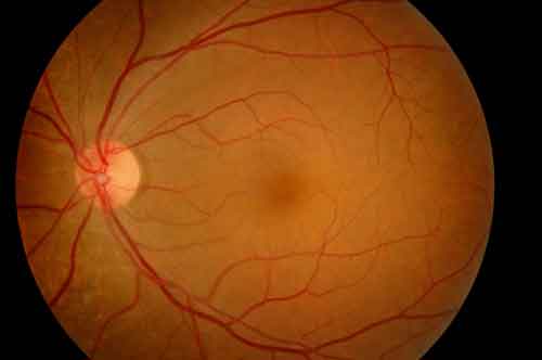
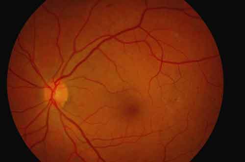
Fundus images requirements
Attention!
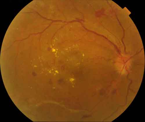
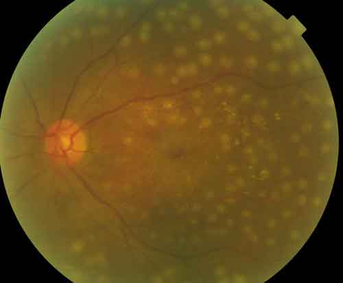
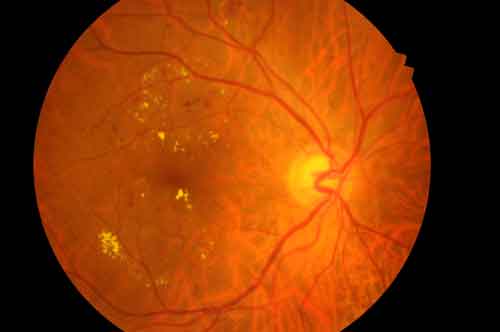
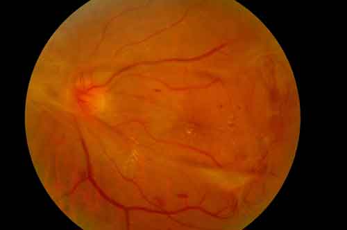
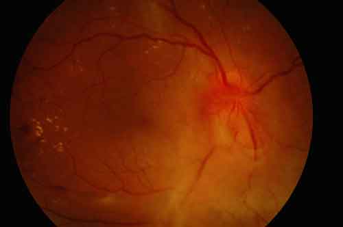
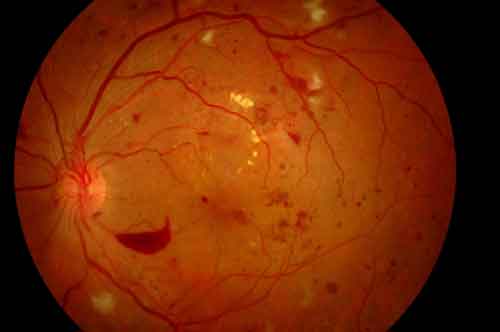
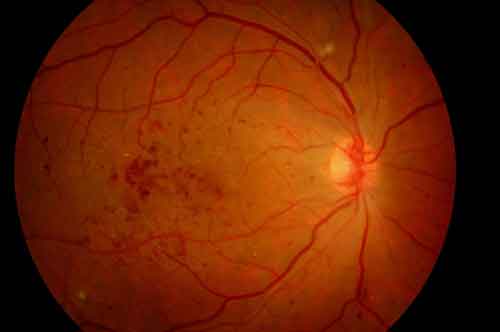
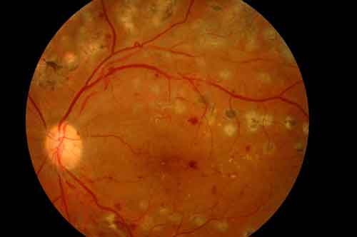
The presence of artifacts in fundus images, poor image quality due to clouding of the optical media, or other reasons can adversely affect the operation of the program and lead to false results. The user is responsible for the quality and compliance with the requirements of images.

Vision-threatening DR includes the following conditions: severe nonproliferative DR, proliferative DR (ETDRS level 53 of greater, but not equal to 90), diabetic macular edema
0
0
0
0

International Council of Ophthalmology Guidelines for Diabetic Eye Care
+ Upload new image